The images presented, comprising various figures and illustrations, were created using BioRender. These figures have been published in The Neuroscientist, a journal that prides itself on making complex neuroscience topics accessible to a wide and diverse readership.
Please note that the current website is in the development phase. To view my established site design, along with various creative projects, feel free to explore Tangible Day.
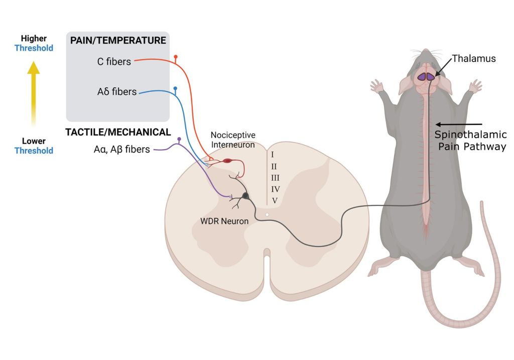
Neural Circuitry of Pain and Tactile Sensation: An Overview of Sensory Fiber Pathways in Rodents. This illustration depicts the neural pathways involved in pain and tactile sensation in a rodent model. It shows different types of sensory fibers: Aα, Aβ fibers for tactile/mechanical sensation with a lower threshold, and C and Aδ fibers for pain/temperature with a higher threshold. These fibers converge on a wide dynamic range (WDR) neuron in the dorsal horn of the spinal cord, which also receives input from a nociceptive interneuron. The WDR neuron then transmits the signal up the spinothalamic pain pathway towards the thalamus in the brain, delineated here in the rodent’s central nervous system. The diagram visually differentiates the pathways by color-coding, suggesting their distinct roles in sensory processing.
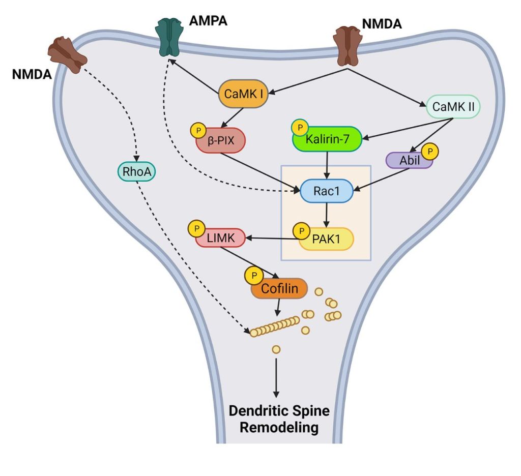
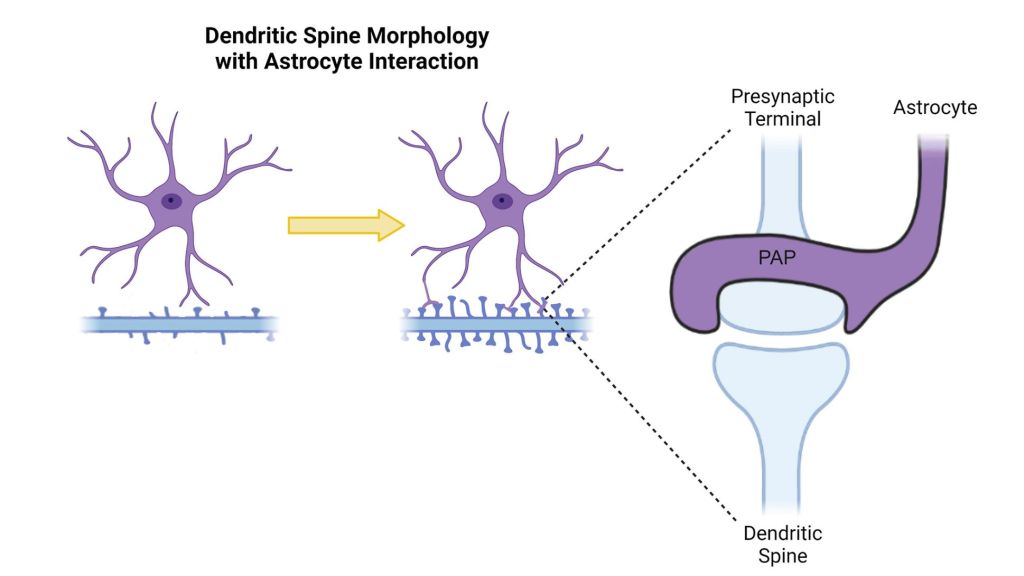
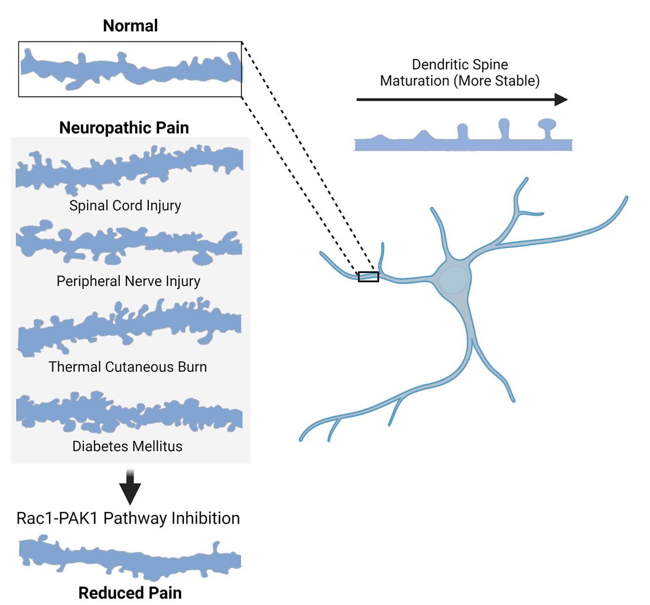
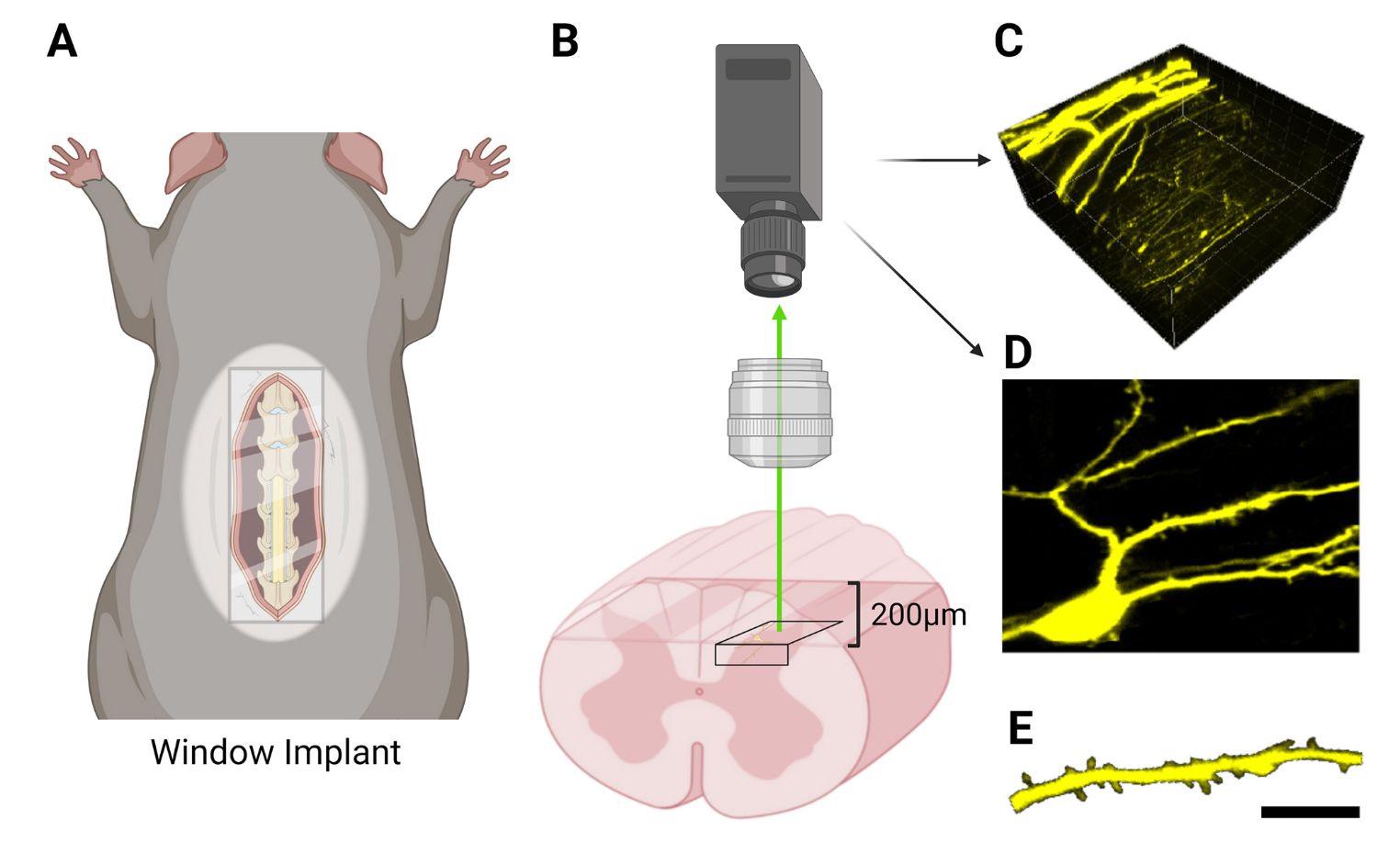
Molecular Dynamics in Dendritic Spines: Rac1 and PAK1 Targets for Drug Development. Proposed molecular cascade within a dendritic spine protrusion on a neuron within the central nervous system (CNS). “Rac1 and PAK1” are molecular targets for potential therapeutic interventions in neurological disease, as they play a key role in the functional organization dendritic spines. Astrocyte Influence on Dendritic Spine Development and Synaptic Integration. This diagram illustrates the interaction between dendritic spine morphology and astrocyte involvement. On the left, a neuron with simple dendritic spines is shown, which, through the process depicted by the arrow, progresses to a more complex spine morphology due to astrocyte interaction, as shown on the right. The close-up view highlights the presynaptic terminal in conjunction with a protruding astrocytic process (PAP), interfacing directly with the dendritic spine, suggesting the astrocyte’s active role in synaptic formation and modulation.Dendritic Spine Plasticity in Neuropathic Pain and the Therapeutic Potential of Rac1-PAK1 Pathway Inhibition. This illustration contrasts the dendritic spine morphology in normal conditions against various neuropathic pain states, including spinal cord injury, peripheral nerve injury, thermal cutaneous burn, and diabetes mellitus. The morphology changes are depicted as pseudo-camera lucida traces. It also suggests that the inhibition of the Rac1-PAK1 signaling pathway can lead to altered dendritic spine morphology and is associated with reduced pain. The arrow pointing downwards indicates the potential therapeutic strategy of targeting this pathway to alleviate pain.In Vivo Imaging Technique for Neuronal Structures in Rodent Spinal Cord. This image series illustrates a process for in vivo imaging of neuronal structures in a rodent model. Panel A shows a rodent with a spinal window implant allowing optical access to dorsal spinal cord tissue. Panel B represents an side view of a microscope objective lens with camera with imaging location into the spinal cord, through the window implant. Panel C displays a three-dimensional reconstruction of the imaged tissue. Panel D provides a high-resolution two-dimensional image of the neuronal structures. Finally, Panel E offers a detailed view of a single dendritic branch, showcasing the level of detail that can be obtained through this imaging technique.
Reference
Benson CA, King JF, Reimer ML, Kauer SD, Waxman SG, Tan AM. Dendritic Spines and Pain Memory. Neuroscientist. 2022 Dec 3:10738584221138251. doi: 10.1177/10738584221138251. Epub ahead of print. PMID: 36461773.

Leave a comment